
PATELLA DISLOCATION
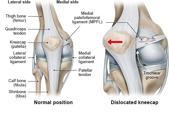
The patella, or kneecap, normally sits in a shallow groove at the end of the thigh bone and glides up and down that groove as the knee bends and straightens. A dislocation happens when it comes out of the groove entirely, almost always slipping towards the outer side of the knee.
It is a common injury in teenagers and young adults, particularly during sport and dance, and it is more common in young women. It is frightening when it happens — the knee often looks visibly wrong — but the immediate injury usually settles, and the more important question is what happens next.
That question matters because a dislocated kneecap has a significant chance of dislocating again, and the risk varies enormously between individuals depending on the shape of the knee. Some people dislocate once and never again. Others have knees anatomically predisposed to it, and for them, recurrence is likely without treatment.
How the kneecap normally stays in place
Three things work together to keep the patella tracking correctly.
The bony groove. The end of the thigh bone has a V-shaped groove — the trochlea — that the patella sits in. A well-formed groove with a prominent outer wall physically prevents the patella escaping sideways.
The ligaments. The most important is the medial patellofemoral ligament, or MPFL, which runs from the inner side of the kneecap to the thigh bone and acts as a checkrein preventing the patella sliding outwards. It is the main soft-tissue restraint in the first 20 to 30 degrees of bending, which is exactly the range in which dislocations occur.
The muscles and alignment. The quadriceps controls the patella actively, and the overall alignment of the leg determines the direction of pull on it. The inner part of the quadriceps, the vastus medialis, is particularly important in resisting outward movement.
A dislocation happens when the forces acting on the patella overcome all three. In a knee with a normal groove and good alignment, that takes a significant injury. In a knee where the groove is shallow or the alignment unfavourable, it can happen with something as ordinary as turning to walk away.
What happens during a dislocation
Two things occur that shape the treatment.
The MPFL tears. In the great majority of first-time dislocations, the medial patellofemoral ligament is torn or stretched as the patella escapes outwards. It has limited capacity to heal back to its original strength, and a stretched or incompetent MPFL is the main reason a knee that has dislocated once is more likely to do so again.
The joint surfaces can be damaged. As the patella dislocates and relocates, its underside strikes the outer edge of the thigh bone. This bruises the bone in almost all cases, and in a meaningful minority chips off a fragment of cartilage and bone — an osteochondral fracture. That fragment becomes a loose body inside the knee.
Identifying an osteochondral fracture is the single most important thing to establish after a first dislocation, because it changes the management completely. A loose fragment that can be fixed back should be fixed promptly, and one that cannot needs removing. Missing it leads to a knee that catches, locks and deteriorates. This is the reason imaging is arranged after a first dislocation even when the knee has settled and feels reasonable.
Why some knees are prone to it
Most people who dislocate a kneecap have some underlying anatomical predisposition. Understanding which factors are present is what allows the risk of it happening again to be estimated with reasonable confidence.
Trochlear dysplasia — a groove that is too shallow, flat, or in some cases convex. This is the strongest single risk factor, associated with roughly three times the risk of recurrence.
Patella alta — a kneecap that sits higher than usual, so it has to travel further before it engages properly in the groove. Associated with around one and a half times the risk.
Skeletal immaturity. Patients whose growth plates are still open have roughly twice the recurrence risk of those who have finished growing.
Age. Risk falls steadily with age — very roughly, each additional year at the time of the first dislocation reduces the recurrence risk by about 8%. A first dislocation at 14 is a very different proposition from a first dislocation at 30.
Alignment, including knock-knee and rotational differences in the thigh or shin bone, and the distance between the groove and the attachment of the patellar tendon.
Generalised ligamentous laxity — being naturally hypermobile.
A dislocation that happened during sport carries about twice the recurrence risk of one that happened during ordinary activity, probably because it reflects the level of demand the knee will be put back under.
The factors compound. A teenager with a shallow groove and a high-riding patella has a substantially higher risk than either factor alone would suggest — reported in the range of 60 to 70% within five years.
Symptoms, and what to do at the time
At the moment of dislocation the knee usually gives way and the patient falls. The kneecap is visibly displaced to the outer side, the knee is held bent, and it is very painful.
The patella often relocates on its own as the knee is straightened, sometimes as the person or a bystander straightens the leg. This is normal and not harmful. Where it does not, it is reduced in an emergency department, usually easily and with pain relief.
Afterwards the knee swells substantially over the following hours — the swelling reflects bleeding into the joint — and is painful, particularly along the inner border of the kneecap where the MPFL has torn. Weight-bearing is difficult initially.
A partial dislocation, or subluxation, is when the patella shifts partly out of the groove and returns on its own. It causes a sharp sensation of the knee giving way, often with pain and swelling afterwards, and is part of the same condition.
After a first dislocation the knee should be assessed and X-rayed, even if it has gone back in and feels better. The purpose is to exclude an osteochondral fracture.
Treatment after a first dislocation
For most first-time dislocations, treatment is non-operative. The exception is where there is an osteochondral fracture or a loose fragment in the joint, which is dealt with surgically and promptly.
Non-operative treatment is a structured rehabilitation programme rather than simply resting.
Early phase. A brace or splint is used for comfort and support initially, with crutches while walking is painful. Ice and elevation settle the swelling. Full immobilisation is avoided beyond the first short period, as stiffness and muscle wasting develop quickly.
Rehabilitation. The focus is on restoring movement and then rebuilding the quadriceps, particularly the inner portion, along with hip and gluteal strength — hip control has a substantial influence on how the knee tracks, and is often the more productive target. Balance and landing mechanics are addressed as activity resumes.
Return to sport is generally around three months, guided by strength and control rather than the calendar.
Physiotherapy makes a real difference here, and a proper programme is worth completing even in someone whose knee feels settled after a few weeks.
When it happens again
Around 30 to 40% of people will dislocate the kneecap again after a first episode. Where a second dislocation occurs, the risk of further episodes rises steeply — to somewhere in the region of 60 to 80%.
Recurrences most often happen within the first year or two, with the average around 16 months after the first episode.
Recurrent instability is not simply a matter of repeated inconvenience. Each dislocation risks further cartilage damage, and repeated episodes contribute to pain, loss of confidence and, over the longer term, arthritis behind the kneecap. Many patients also develop a persistent apprehension about the knee that limits what they will do, even between episodes.
Surgery is generally recommended after a second dislocation, and is considered earlier in patients with strongly unfavourable anatomy or with an osteochondral injury.
Surgery for recurrent instability
Surgery aims to restore the restraint that has been lost and, where necessary, to correct the underlying anatomy. The procedure is tailored to what the assessment shows rather than being one standard operation.
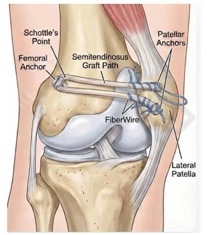
MPFL reconstruction is the mainstay. A new ligament is created, usually using one of the patient's own hamstring tendons, running from the inner border of the patella to the correct point on the thigh bone, reproducing the original ligament. It restores the checkrein that prevents the patella sliding outwards. For most patients with recurrent instability and reasonable underlying anatomy, this alone is sufficient.
Tibial tubercle osteotomy is added where the alignment is a significant contributor. The bony prominence where the patellar tendon attaches to the shin bone is detached, repositioned and fixed with screws, changing the direction of pull on the patella and, where needed, lowering a high-riding patella. It adds bone healing time to the recovery.
Loose fragments are addressed at the same time, fixed back where possible or removed where not.
Results are good. Recurrent dislocation after surgery occurs in fewer than 10% of patients, and most return to their previous activities.
Recovery after surgery
Surgery is usually performed as a day case. For the first two weeks, a brace is worn, and crutches may be used. Gentle movement exercises begin early to prevent stiffness. Pain settles over the first week or two.
Once the brace is removed, range of movement and quadriceps strength progress under physiotherapy guidance commences. Driving typically becomes possible somewhere between four and six weeks, once off crutches and able to control the car confidently.
Running is generally resumed at around three months. Following this, sport-specific work — agility, cutting, jumping and landing. Return to competitive sport is usually around six months, based on meeting strength and control benchmarks rather than the date alone.
Risks and complications
Surgery for patellar instability is well established and serious complications are uncommon.
Infection, which is uncommon and usually settles with antibiotics; deep infection occasionally requires further surgery.
Blood clots in the leg or lung, which are uncommon but recognised.
Stiffness, particularly loss of bending, which is the most common issue and is minimised by starting movement early.
Delayed or incomplete bone healing where a tibial tubercle osteotomy has been performed, occasionally requiring further treatment. The screws sometimes become prominent and irritating, and are removed in a small number of patients.
Ongoing pain at the front of the knee, which can persist even where the instability has been successfully corrected.
Further dislocation, in fewer than 10% of patients.
Fracture of the patella, which is rare but recognised where tunnels are made in the kneecap.
Numbness around the incisions, which is common and rarely troublesome.
ACC and costs
When the patellar dislocation is a traumatic injury, treatment is generally covered by ACC.
Where the underlying cause is anatomical, the position can occasionally be less straightforward, and claims are assessed individually.