
CALCIFIC TENDONITIS
Calcific tendonitis is a condition in which a deposit of calcium forms inside one of the rotator cuff tendons of the shoulder. It can be entirely painless for years, and then, over a period of days, become one of the most severe pains encountered in orthopaedics.
Two things are worth knowing at the outset, because they change how the condition feels to live with.
The first is that it is not arthritis, and it is not a tear. The tendon is not wearing out or coming apart. A deposit has formed within it, and the body's attempt to clear that deposit is what produces the pain.
The second is that it is usually self-limiting. In most people the deposit is eventually resorbed and the shoulder returns to normal, often completely. Treatment is largely about controlling symptoms and, where appropriate, speeding that process along — not about repairing damage.
What calcific tendonitis is
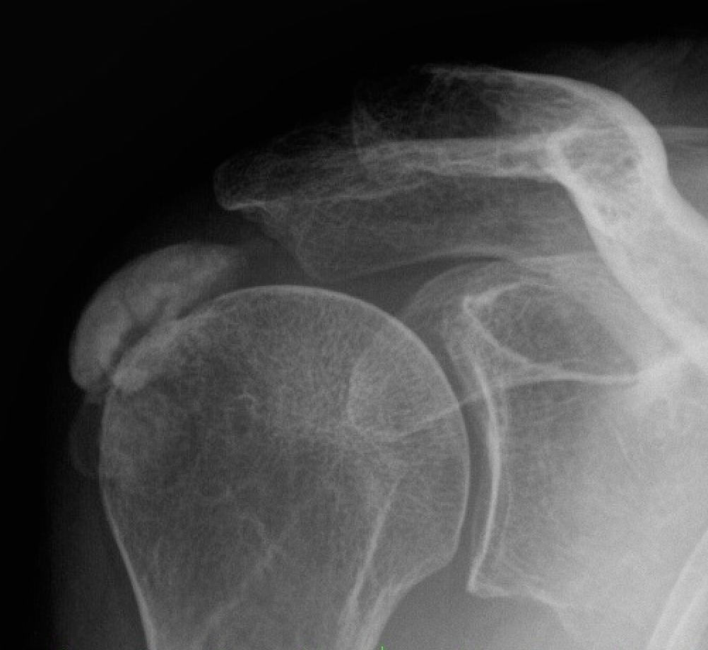
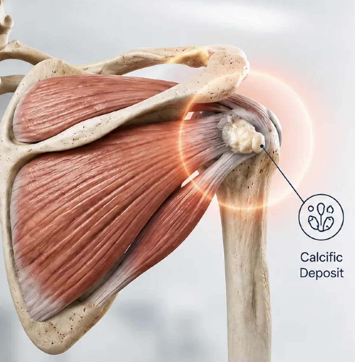
The rotator cuff is a group of four muscles and tendons that wrap around the shoulder joint, holding the ball centred in its socket and powering movement of the arm. In calcific tendonitis, a deposit of calcium hydroxyapatite — chemically similar to the mineral in bone, but chalky and paste-like rather than solid — forms within the substance of one of these tendons. The supraspinatus tendon, at the top of the shoulder, is much the most common site.
The deposit itself is not the direct cause of pain in most cases. Many people have one and never know. Pain arises for two reasons: a large deposit can raise pressure inside the tendon and take up space beneath the bony arch of the shoulder, causing a persistent ache with movement; and when the body begins to break the deposit down, it mounts an intense inflammatory response to clear it, which is what produces the severe acute pain that brings most people to seek help.
That second point explains something patients find counter-intuitive, and it is worth stating plainly: the most painful phase is the phase in which the problem is resolving.
Who gets it, and why
Calcific tendonitis most often affects people between about 30 and 60, and is more common in women. It is common enough that calcium deposits are found in roughly 8% of shoulders in people with no symptoms at all, and in a much higher proportion of those presenting with shoulder pain.
The underlying cause is not fully understood. The leading explanation is that a localised area of tendon undergoes a change in cell type and begins to deposit calcium, possibly triggered by a reduction in local blood supply or by low-grade tendon stress. Diabetes and thyroid disorders are associated with a higher incidence and with a more prolonged course.
Two common assumptions are worth correcting directly:
It has nothing to do with dietary calcium. Calcium intake, dairy consumption, calcium supplements and blood calcium levels are all unrelated to this condition. Reducing calcium in the diet will not help and is not advised.
It is not caused by an injury or by overuse. It is not something a patient did to themselves. It can occur in a shoulder that has never been injured and in people who do no repetitive or overhead work at all.
The stages, and why they matter
Calcific tendonitis follows a recognised sequence. Knowing which stage a shoulder is in explains the symptoms and, importantly, guides what treatment makes sense.
Pre-calcific stage. Tendon tissue undergoes changes that predispose it to forming a deposit. There are no symptoms.
Formative stage. Calcium is laid down within the tendon. The deposit is firm and chalky. This stage is often painless, or causes only a mild ache with overhead activity. It can last months or years.
Resting stage. The deposit is stable and quiet. Many people remain in this phase indefinitely and never develop significant symptoms. A deposit found incidentally on an X-ray taken for another reason is usually in this state.
Resorptive stage. The body begins to break the deposit down and clear it. Blood vessels grow into the area and inflammatory cells arrive to remove the calcium, which softens to a toothpaste-like consistency and can leak into the surrounding space. This is the acutely painful phase — and it is the phase from which the condition resolves.
Post-calcific stage. The deposit has gone and the tendon remodels back towards normal tissue. Pain settles and function returns, usually completely.
The practical implication is significant. A patient in the middle of the resorptive phase is in severe pain but is also, in a real sense, getting better. Deposits in this phase have the highest rate of spontaneous resolution — a majority clear within a few years without any intervention — and treatment during it is aimed at making the process bearable rather than at rescuing a deteriorating situation.
Symptoms
Calcific tendonitis presents in two quite different ways.
The chronic pattern. A persistent ache in the shoulder, worse with overhead activity and often worse at night, sometimes with a catching sensation as the arm is raised. This can grumble along for months and is easily mistaken for other rotator cuff problems.
The acute pattern. Pain that escalates over hours to days to become severe and constant, present at rest, unrelieved by position, and preventing sleep entirely. The shoulder is often held rigidly still because any movement is intolerable. Patients frequently describe it as among the worst pain they have experienced, and it is not unusual for people to present to an emergency department.
The acute pattern is alarming precisely because it appears without warning and without injury. It typically peaks over one to two weeks and then settles over the following few weeks as the deposit clears.
Treatment
Treatment is guided by which stage the shoulder is in and by how severe the symptoms are. The great majority of patients never need surgery.
Reassurance and time
For a deposit found incidentally, or one causing only mild symptoms, no active treatment is required. Deposits frequently resolve on their own — and are more likely to do so once the resorptive phase has begun.
For a patient in an acute episode, understanding what is happening genuinely helps. The pain is severe but time-limited, the shoulder is not being damaged, and the process is self-resolving.
Pain relief
During an acute episode, effective pain control is the priority. Anti-inflammatory medication is the mainstay and is often taken regularly for a short period rather than only as needed. Paracetamol is added for background relief. Occasionally a short course of stronger pain relief is appropriate, and in a severe acute episode this is entirely reasonable — the pain is genuinely severe and there is no benefit in enduring it.
Ice can help during the acute phase. Supporting the arm in a sling for short periods may make an acute episode more tolerable, though prolonged immobilisation is avoided because of the risk of secondary stiffness.
Physiotherapy
Physiotherapy has a limited role during a severe acute episode, when the shoulder is too painful to move usefully. It becomes valuable once pain begins to settle, and its main purposes are to restore range of movement, prevent the shoulder stiffening up, and rebuild strength around the shoulder blade and rotator cuff after a period of disuse.
Preventing stiffness matters. A significant proportion of the ongoing disability after calcific tendonitis comes not from the deposit but from a shoulder that was held still for too long.
Corticosteroid injection
An injection of corticosteroid into the subacromial space — the space above the tendon — does not dissolve the deposit, but it can substantially reduce the inflammation surrounding it and provide meaningful relief during an acute episode. It is often what makes the difference between a manageable few weeks and an unmanageable one, and it allows physiotherapy to begin.
Studies of physiotherapy, anti-inflammatories and subacromial injection together report clinical improvement in roughly three-quarters of patients.
Ultrasound-guided barbotage (needling and lavage)
Barbotage is a procedure performed under ultrasound guidance and local anaesthetic, usually taking around twenty minutes. A needle is passed into the deposit and saline is injected and withdrawn repeatedly, breaking the calcium up and washing it out. A corticosteroid injection into the subacromial space is generally given at the same time.
It is the most direct non-surgical treatment available, and the evidence supporting it is reasonably good. Studies report calcium resorption in around three-quarters of treated shoulders, compared with roughly 40% of untreated controls, and combining barbotage with a steroid injection roughly doubles the rate of complete resolution at one year compared with a steroid injection alone.
It is most suitable for deposits that are softening or in the resorptive phase, as these can be aspirated more readily. A firm, dense deposit may not break up as easily. Some increase in pain for a day or two afterwards is common, and patients are warned about this so it is not mistaken for a complication.
Extracorporeal shockwave therapy
Shockwave therapy delivers focused acoustic energy through the skin to the deposit, over a course of sessions. It is thought to work by fragmenting the calcium and stimulating the local blood supply and resorptive response.
The evidence favours high-energy focused treatment specifically: high-energy shockwave therapy has been reported to clear the deposit in a substantial majority of patients, with good clinical results maintained at one year, whereas low-energy treatment performs little better than placebo. Where it has been compared directly with ultrasound-guided barbotage, barbotage has generally cleared deposits more reliably, though both improve symptoms.
It is non-invasive, requires no needle, and is a reasonable option for patients who prefer to avoid a procedure. Treatment is uncomfortable, and several sessions are usually required.
Surgery
Surgery is reserved for the minority of patients whose symptoms persist despite the measures above — typically someone with a large, persistent deposit, ongoing significant pain over many months, and no response to injection or barbotage.
Results are good, with around three-quarters to four-fifths of patients reporting a satisfactory outcome. Temporary stiffness afterwards is the most common issue, affecting a proportion of patients and usually resolving with physiotherapy.
Risks include the general risks of surgery and anaesthesia, which occur in less than 1% of cases, and infection, at approximately 1%. Other recognised risks are stiffness, incomplete removal of the deposit with persisting symptoms, and, uncommonly, the need for further surgery.