
KNEE ARTHRITIS
Knee arthritis is wear of the smooth cartilage lining the knee joint. As that lining thins, the bones begin to rub against one another, and the knee becomes painful, stiff and often swollen. It is the most common form of arthritis affecting a major joint, and it develops gradually over years.
The important thing to say at the outset is that people with knee arthritis can be managed successfully without surgery, often for many years, and that the treatments with the strongest evidence behind them are things a patient can start on themselves.
A second point worth making early, because it causes a great deal of unnecessary worry: being told a knee is "bone on bone" is not a diagnosis that requires surgery. It describes what an X-ray shows. Plenty of people with that appearance manage well for years, and plenty of people with mild-looking X-rays have significant symptoms. What matters is how the knee behaves, not how it looks on x-ray.
Understanding the knee joint
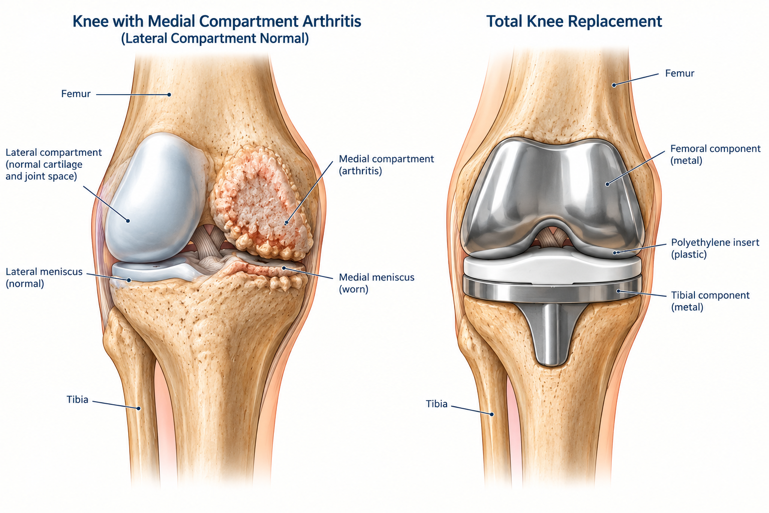
The knee is where the thigh bone (femur), the shin bone (tibia) and the kneecap (patella) meet. It is usually described as having three compartments: the medial compartment on the inner side, the lateral compartment on the outer side, and the patellofemoral compartment where the kneecap runs in its groove at the front.
The ends of the bones are coated in articular cartilage, a firm, slippery layer a few millimetres thick that allows the joint to glide almost without friction. Between the femur and tibia sit two crescent-shaped menisci, which act as shock absorbers and help spread load across the joint. Ligaments — including the cruciate ligaments in the centre — hold the knee stable, and the surrounding muscles, particularly the quadriceps, control and protect it.
In osteoarthritis, the cartilage gradually wears away. As it does, several things happen together: the joint space narrows, bone spurs (osteophytes) form around the edges, the lining of the joint becomes inflamed and produces excess fluid, and the bone underneath the worn cartilage thickens and becomes tender. The menisci often degenerate at the same time.
Wear is frequently uneven. The medial compartment is affected most often, and as that side wears down the leg gradually bows inwards — a knee that becomes progressively more bow-legged over years is usually telling you where the wear is.
What causes knee arthritis?
Osteoarthritis is usually the result of several factors acting together rather than any single cause.
Age. The strongest single factor. Cartilage becomes less resilient over time, and arthritis becomes considerably more common beyond the age of 50.
Genetics. A family history meaningfully increases risk. Some people simply have cartilage that wears earlier, and this cannot be changed.
Body weight. The knee carries several times body weight with each step, so additional weight has an amplified effect on the joint. Weight also contributes through low-grade inflammation, which is why it affects joints that are not weight-bearing as well. This is one of the few risk factors that can be modified, which is why it comes up in almost every consultation.
Previous injury. Arthritis following an injury is called post-traumatic arthritis. Ligament injuries — particularly ACL ruptures, meniscal tears and fractures involving the joint surface all increase the risk, sometimes decades later. This is a common route to arthritis in younger patients.
Previous knee surgery, particularly removal of a significant portion of a meniscus, which reduces the knee's ability to spread load.
Alignment. Bow-legged or knock-kneed alignment concentrates load on one side of the joint and accelerates wear there.
Occupation and activity. Work involving heavy lifting, prolonged kneeling or squatting is associated with a higher risk. Ordinary recreational exercise, including running in people with healthy knees, is not.
Symptoms
Knee arthritis typically causes pain that is worse with weight-bearing activity and better with rest, at least in the earlier stages. Stairs, particularly going down, and getting up from a low chair are often the first things to become difficult.
Stiffness is common first thing in the morning and after sitting for a while, and usually eases within about half an hour of moving. Stiffness that lasts substantially longer than that, or that affects many joints, raises the possibility of an inflammatory arthritis rather than osteoarthritis.
Swelling comes and goes, often after activity, and can make the knee feel tight and heavy.
Grinding, clicking or catching is common and is not in itself a sign of damage progressing.
Giving way may occur, usually because the quadriceps muscle has weakened rather than because the knee is unstable.
Night pain and pain at rest tend to appear later, and are among the symptoms that most reliably indicate the arthritis has become significant.
The pattern is typically gradual over years, with flares lasting weeks to months separated by quieter periods. A flare is not evidence of permanent deterioration.
What to expect over time
Osteoarthritis is generally slowly progressive, but the rate varies enormously and cannot be predicted from a first X-ray. Some knees change very little over a decade. Symptoms fluctuate substantially, and most patients have periods that are considerably better and worse than their average.
Well-managed knee arthritis often stays at a manageable level for many years. Delaying surgery in a well-managed knee does not usually compromise the eventual result, so there is no pressure to make decisions quickly.
Non-surgical treatment
The goals are to control pain, keep the knee moving, maintain the strength that protects it, and preserve independence in the activities that matter. These are achievable for the great majority of people.
No treatment currently available regrows cartilage or reverses the underlying arthritis. What follows is aimed at symptoms and function — which is what determines quality of life.
It is worth knowing that the two treatments with the strongest evidence behind them, by some distance, are exercise and weight management. Both are things a patient controls directly. Neither is a consolation prize offered while waiting for surgery.
Exercise and strengthening
Exercise is the single most effective non-operative treatment for knee arthritis, and it is recommended as a core treatment by every major clinical guideline. It reduces pain and improves function, with effects comparable to those of anti-inflammatory medication and without the side effects.
The mechanism is straightforward. A strong quadriceps muscle absorbs load that would otherwise pass through the worn joint surfaces, and it controls the knee through movement. Weak quadriceps is both a consequence of knee arthritis and a cause of its progression, and breaking that cycle changes how the knee feels.
A useful programme has three elements:
Strengthening, particularly of the quadriceps but also the hamstrings and hip muscles. Straight-leg raises, sit-to-stand repetitions, wall squats within a comfortable range, step-ups and resistance band work are typical. This is the part that matters most.
Low-impact aerobic exercise to maintain fitness and assist with weight management. Stationary cycling is particularly well suited to arthritic knees — the load through the joint is low and the movement helps with stiffness. Swimming, aqua-jogging and walking on level ground are all appropriate.
Range of movement work, especially maintaining the ability to straighten the knee fully, which is easily lost and affects walking.
Two things are worth being clear about. First, some discomfort during and after exercise is expected and acceptable.Pain that settles within an hour or so is not a sign of harm. Pain that is sharply worse and persists into the next day means the load was too high — ease the programme, rather than abandoning it. Second, this takes time. Meaningful improvement typically takes six to twelve weeks of consistent work, and the benefit fades if the programme stops.
Weight management
Where weight is a factor, this is the intervention with the largest effect on symptoms after exercise.
The reason is mechanical as well as metabolic. The knee carries roughly four times body weight with each step during normal walking, so each kilogram lost removes something in the order of four kilograms of load from the knee with every step taken. Over a day's walking, that is a substantial change in what the joint has to absorb. Weight also drives low-grade inflammation, which contributes independently.
The evidence suggests that more is better: a loss of around 10% of body weight produces considerably greater improvement in pain and function than a loss of 5%, and combining weight loss with exercise works better than either on its own.
Activity modification
Adjusting how the knee is used — rather than using it less — is often the change that makes the most practical difference.
The load through the knee rises steeply with stairs, squatting, kneeling, and carrying heavy loads. Practical adaptations include taking stairs one at a time leading with the better leg going up and the worse leg going down, using a handrail, sitting on a stool rather than kneeling for garden or floor work, breaking heavy tasks into shorter blocks, carrying loads in two lighter trips, and choosing supportive cushioned footwear.
Switching some walking to cycling or swimming maintains fitness while reducing joint load, and is often more sustainable than trying to push through walking that has become painful.
Walking aids and braces
A walking stick, held in the hand opposite the affected knee, meaningfully reduces load through the joint and is worth trying during flares or for longer distances. Many patients resist this, and it is worth saying that using a stick when it helps generally allows people to stay more active, not less.
Braces and sleeves help some patients. A simple elastic sleeve provides warmth and a sense of support and is inexpensive to trial. An offloading brace, designed to shift load away from the worn compartment, can be useful in selected patients with wear confined to one side, though they are bulky and not everyone tolerates them.
Footwear and insoles. Cushioned, supportive shoes reduce impact. Specific orthoses have less consistent evidence but are reasonable to trial.
Topical anti-inflammatory gels
Topical anti-inflammatory gel applied over the knee is a good first choice for medication. Because the knee is a relatively superficial joint, topical preparations work well here — guidelines regard them as comparable in effectiveness to oral anti-inflammatories for the knee, with far less absorption into the body and correspondingly lower risk. For many patients this is all that is needed.
Simple pain relief
Paracetamol taken regularly — rather than only when pain is bad — remains a reasonable option and is well tolerated by most people. The usual maximum is 4 grams in 24 hours for adults, and lower in some circumstances. Its effect in knee arthritis is modest, and it works best as background relief alongside other measures.
Oral anti-inflammatory medications are more effective, particularly during a flare. They carry gastrointestinal, kidney and cardiovascular risks and are best used at the lowest effective dose for short periods rather than continuously. A useful approach is to take them around specific demanding activities rather than every day. Anyone with existing heart, kidney, blood pressure or stomach problems, or taking other regular medications, should discuss them with their GP or pharmacist first.
Opioid pain medications have no useful role in the long-term management of knee arthritis. They perform poorly in this condition, carry significant risks, and are not recommended.
Corticosteroid injection
An injection of corticosteroid into the knee can settle a painful flare and is most useful when pain is severe enough to prevent someone doing their exercises — it creates a window in which rehabilitation becomes possible.
The relief is real but temporary, commonly a few weeks to a few months, and injections do not slow the underlying arthritis. Repeated frequent injections into the same knee are avoided; there is no fixed limit, but if a knee is needing them regularly the overall plan is worth revisiting. An injection is generally avoided within a few months before a planned knee replacement because of an increase in infection risk.
Other injections
Hyaluronic acid ("gel" injections or viscosupplementation) has been used for many years. The evidence is mixed and the average benefit small; several major guidelines no longer recommend it, while others regard it as of uncertain value. It is not publicly funded.
Platelet-rich plasma (PRP) is widely marketed. Trial results are inconsistent, and the better-quality recent trials have been less encouraging than earlier ones.
Stem cell and similar regenerative therapies are heavily promoted for knee arthritis. They are expensive, and not supported by good evidence at this time. No injection currently available regrows cartilage, whatever the marketing claims.
Supplements
Glucosamine, chondroitin, fish oil and green-lipped mussel extract are frequently asked about. The evidence for symptom relief is weak and inconsistent, and there is no good evidence that any of them slows the progression of arthritis. They are safe for most people to trial, and some patients report benefit. If there is no clear improvement after two to three months, there is little reason to continue.
Heat, ice and simple measures
Heat before activity and ice after are simple, safe and genuinely helpful for many people. Ice is particularly useful for a swollen knee after a demanding day. Attention to sleeping position — a pillow between or under the knees, though not one that holds the knee bent for long periods — can help night discomfort.
What is not recommended
Two things are worth stating plainly, because patients frequently ask about both.
Arthroscopic ("keyhole") surgery for arthritis. This was once common and is now strongly recommended against by clinical guidelines for degenerative knee disease. Multiple high-quality trials have shown that arthroscopic washout, debridement or trimming of a degenerate meniscal tear provides no meaningful benefit over non-operative treatment in an arthritic knee, and carries the risks of an operation and an anaesthetic. A degenerate meniscal tear found on MRI in an arthritic knee is part of the arthritis, not a separate fixable problem.
There are exceptions. A genuinely locked knee, where a torn fragment is physically blocking movement, is a different situation. So is a younger patient with a traumatic tear and little or no arthritis.
Prolonged rest. Avoiding activity leads to weaker muscles, more stiffness and a knee that copes less well. Activity should be modified, not abandoned.
When surgery is considered
Non-operative treatment continues for as long as it is working, and for many people that is indefinitely. Surgery becomes a reasonable discussion when three things are true together: pain and stiffness significantly limit daily life or sleep, a genuine trial of the measures above has not given adequate benefit, and the patient themselves wants an operation.
There is no fixed threshold and no need to rush. Age alone is not a barrier in either direction.
If that point is reached knee replacement— total or, in selected patients, partial — is a reliable operation with well-documented long-term results.
For a smaller group, usually younger patients with wear confined to one compartment and a bow-legged or knock-kneed alignment, an osteotomy may be an option. This realigns the leg to shift load away from the worn side, preserving the patient's own joint and deferring a replacement. It is a bigger recovery than a replacement and suits a specific group, but for the right patient it is a valuable alternative.